Crystallography (2025)")
X-ray Diffraction (XRD) Crystallography: Revealing the Hidden Architecture of Matter. Discover How This Pioneering Technique Transforms Science, Industry, and Innovation. (2025)
- Introduction to X-ray Diffraction (XRD) Crystallography
- Historical Milestones and Nobel-Winning Discoveries
- Principles of XRD: How It Works
- Instrumentation and Technological Advances
- Applications Across Science and Industry
- Case Studies: Breakthroughs Enabled by XRD
- Current Market Trends and Public Interest (Estimated 8% Annual Growth, 2024–2029)
- Key Players and Official Resources (e.g., Bruker.com, Rigaku.com, IUCr.org)
- Challenges, Limitations, and Evolving Solutions
- Future Outlook: Innovations and Expanding Frontiers in XRD Crystallography
- Sources & References
Introduction to X-ray Diffraction (XRD) Crystallography
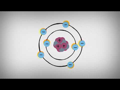
X-ray Diffraction (XRD) Crystallography is a cornerstone analytical technique in materials science, chemistry, geology, and biology, enabling the precise determination of the atomic and molecular structure of crystalline materials. The method is based on the interaction of X-rays with the periodic lattice of a crystal, producing a diffraction pattern that can be mathematically interpreted to reveal the arrangement of atoms within the crystal. Since its inception in the early 20th century, XRD has played a pivotal role in scientific advancements, including the elucidation of DNA’s double helix structure and the development of novel materials.
The fundamental principle underlying XRD crystallography is Bragg’s Law, which relates the wavelength of incident X-rays and the angle at which they are diffracted by the crystal lattice planes. When a monochromatic X-ray beam strikes a crystalline sample, constructive interference occurs at specific angles, resulting in a series of diffracted beams. By measuring the intensities and angles of these beams, researchers can reconstruct a three-dimensional electron density map of the crystal, from which atomic positions are inferred.
Modern XRD instrumentation typically consists of an X-ray source, a goniometer to precisely orient the sample, and a detector to record the diffracted beams. Advances in detector technology, automation, and data analysis software have significantly increased the speed and accuracy of XRD measurements. Laboratories and research facilities worldwide, including those operated by organizations such as the International Union of Crystallography (IUCr), have established standardized protocols and databases to facilitate the sharing and interpretation of crystallographic data.
XRD crystallography is indispensable for characterizing the phase composition, crystallinity, and structural defects of materials. It is widely used in the identification of minerals, the development of pharmaceuticals, the design of advanced functional materials, and the study of biological macromolecules. The technique is also central to quality control in industrial processes, forensic investigations, and the certification of reference materials by standards organizations such as the National Institute of Standards and Technology (NIST).
As of 2025, XRD crystallography continues to evolve, with innovations in synchrotron radiation sources, microfocus X-ray beams, and computational methods expanding its capabilities. The technique remains a vital tool for both fundamental research and applied science, underpinning discoveries across a broad spectrum of disciplines.
Historical Milestones and Nobel-Winning Discoveries
X-ray diffraction (XRD) crystallography has a rich history marked by groundbreaking discoveries and Nobel Prize-winning achievements that have profoundly shaped modern science. The technique’s origins trace back to the early 20th century, when German physicist Max von Laue first demonstrated the diffraction of X-rays by crystals in 1912. This pivotal experiment provided direct evidence of the wave nature of X-rays and the periodic atomic structure of crystals, earning von Laue the Nobel Prize in Physics in 1914. His work laid the foundation for the systematic study of crystal structures using X-ray beams.
Building on von Laue’s discovery, father-and-son team William Henry Bragg and William Lawrence Bragg developed the mathematical framework—now known as Bragg’s Law—that relates the angles at which X-rays are diffracted to the distances between atomic planes in a crystal. Their collaborative efforts enabled the determination of atomic arrangements in solids, a breakthrough that garnered them the Nobel Prize in Physics in 1915. The Braggs’ contributions established XRD crystallography as a powerful tool for elucidating the three-dimensional structures of matter.
Throughout the 20th century, XRD crystallography continued to drive scientific progress. In 1953, Rosalind Franklin’s X-ray diffraction images of DNA, particularly the famous “Photo 51,” were instrumental in revealing the double-helix structure of DNA. This discovery, interpreted by James Watson and Francis Crick, revolutionized molecular biology and led to the awarding of the Nobel Prize in Physiology or Medicine in 1962 to Watson, Crick, and Maurice Wilkins. The role of XRD in this achievement underscored its significance in understanding the molecular basis of life.
Further Nobel Prizes have recognized advances in XRD crystallography. Dorothy Crowfoot Hodgkin received the Nobel Prize in Chemistry in 1964 for her work in determining the structures of important biomolecules, including penicillin and vitamin B12, using X-ray crystallography. More recently, in 2009, the Nobel Prize in Chemistry was awarded to Venkatraman Ramakrishnan, Thomas A. Steitz, and Ada E. Yonath for their studies of the structure and function of the ribosome, again utilizing X-ray crystallography.
Today, XRD crystallography remains a cornerstone of structural science, with organizations such as the International Union of Crystallography and the National Institute of Standards and Technology supporting research, standardization, and education in the field. The technique’s historical milestones and Nobel-winning discoveries continue to inspire innovation across chemistry, biology, materials science, and beyond.
Principles of XRD: How It Works
X-ray Diffraction (XRD) crystallography is a cornerstone analytical technique in materials science, chemistry, geology, and biology, enabling the determination of the atomic and molecular structure of crystalline materials. The fundamental principle of XRD is based on the interaction between incident X-rays and the periodic atomic planes within a crystal lattice. When a monochromatic X-ray beam strikes a crystalline sample, the atoms within the crystal cause the X-rays to scatter in specific directions. This scattering is governed by Bragg’s Law, which relates the wavelength of the X-rays and the angle at which they are diffracted to the distance between the crystal planes.
Bragg’s Law is mathematically expressed as nλ = 2d sinθ, where n is an integer (the order of reflection), λ is the wavelength of the incident X-ray, d is the distance between atomic planes in the crystal, and θ is the angle of incidence at which constructive interference occurs. Constructive interference leads to the formation of distinct diffraction peaks, which are detected and recorded as a diffraction pattern. Each crystalline substance produces a unique pattern, serving as a “fingerprint” for phase identification and structural analysis.
The process of XRD crystallography typically involves several key steps. First, a finely powdered or single-crystal sample is prepared and mounted in the path of an X-ray beam. As the X-rays interact with the sample, a detector measures the intensity and angles of the diffracted beams. The resulting data are plotted as intensity versus angle (2θ), producing a diffraction pattern. By analyzing the positions and intensities of the peaks, researchers can deduce the crystal structure, lattice parameters, and even the arrangement of atoms within the unit cell.
Modern XRD instruments employ sophisticated X-ray sources, such as sealed tubes or synchrotron radiation, and highly sensitive detectors to enhance resolution and data quality. The technique is non-destructive and applicable to a wide range of materials, including metals, minerals, polymers, and biological macromolecules. XRD is also fundamental in the determination of unknown compounds, quality control, and the study of phase transitions.
Globally, organizations such as the International Union of Crystallography (IUCr) play a pivotal role in advancing the science of crystallography, setting standards, and fostering collaboration among researchers. The National Institute of Standards and Technology (NIST) also provides reference materials and databases crucial for XRD analysis. These authoritative bodies ensure the reliability and reproducibility of XRD methodologies, supporting its continued evolution as a vital tool in scientific research.
Instrumentation and Technological Advances
X-ray Diffraction (XRD) crystallography has undergone significant technological evolution, driven by advances in instrumentation and analytical methodologies. At its core, XRD relies on the interaction of X-rays with the periodic atomic planes in crystalline materials, producing diffraction patterns that reveal structural information. The precision and efficiency of this technique are fundamentally linked to the quality and sophistication of the instrumentation employed.
Modern XRD instruments are characterized by high-brilliance X-ray sources, advanced optics, sensitive detectors, and robust computational capabilities. The transition from traditional sealed-tube X-ray sources to microfocus and rotating anode generators has markedly increased X-ray intensity, enabling faster data collection and improved resolution. Furthermore, the integration of monochromators and advanced collimation systems has enhanced beam quality, reducing background noise and improving signal-to-noise ratios.
A major leap in XRD technology has been the widespread adoption of two-dimensional (2D) and hybrid pixel detectors. These detectors, such as those based on silicon or cadmium telluride, offer rapid readout speeds, high dynamic range, and low noise, facilitating the collection of high-quality diffraction data even from minute or weakly diffracting samples. The use of area detectors has also enabled the development of high-throughput screening and in situ experiments, expanding the applicability of XRD in fields such as pharmaceuticals, materials science, and catalysis.
Automation and robotics have further transformed XRD crystallography. Automated sample changers, robotic arms, and integrated software platforms now allow for unattended, high-throughput data acquisition and analysis. These systems are particularly valuable in industrial and academic settings where large numbers of samples must be processed efficiently. The implementation of artificial intelligence and machine learning algorithms in data processing pipelines has accelerated structure solution and refinement, reducing human intervention and minimizing errors.
Synchrotron radiation facilities, such as those operated by European Synchrotron Radiation Facility and Advanced Photon Source, have played a pivotal role in pushing the boundaries of XRD. These large-scale research infrastructures provide extremely intense and tunable X-ray beams, enabling studies of very small crystals, time-resolved processes, and complex biological macromolecules. The development of free-electron lasers and next-generation synchrotrons promises even greater temporal and spatial resolution, opening new frontiers in crystallographic research.
In summary, the instrumentation and technological advances in XRD crystallography as of 2025 have greatly expanded the technique’s capabilities, making it faster, more sensitive, and more versatile. These innovations continue to drive discoveries across chemistry, biology, physics, and materials science, cementing XRD’s role as a cornerstone of structural analysis.
Applications Across Science and Industry
X-ray Diffraction (XRD) crystallography is a cornerstone analytical technique with wide-ranging applications across science and industry. Its fundamental ability to elucidate the atomic and molecular structure of crystalline materials has made it indispensable in fields such as materials science, chemistry, geology, pharmaceuticals, and engineering. By analyzing the diffraction patterns produced when X-rays interact with a crystal lattice, XRD provides detailed information about unit cell dimensions, atomic positions, and the overall arrangement of atoms within a material.
In materials science, XRD is routinely used to identify phases, determine crystallite size, and assess the degree of crystallinity in metals, ceramics, polymers, and composites. This information is critical for tailoring material properties for specific applications, such as improving mechanical strength, thermal stability, or corrosion resistance. For example, the development of advanced alloys and high-performance ceramics often relies on XRD analysis to monitor phase transformations and optimize processing conditions.
The pharmaceutical industry leverages XRD crystallography to characterize active pharmaceutical ingredients (APIs) and excipients, ensuring the correct polymorphic form is present for optimal drug efficacy and stability. Regulatory agencies, such as the U.S. Food and Drug Administration, recognize XRD as a key tool for verifying the identity and purity of pharmaceutical compounds. XRD also plays a vital role in the discovery and development of new drugs by enabling the determination of protein-ligand structures, which informs rational drug design.
In geology and mineralogy, XRD is essential for the identification and quantification of minerals in rocks, soils, and sediments. Organizations like the U.S. Geological Survey employ XRD to analyze mineral compositions, which aids in resource exploration, environmental monitoring, and understanding geological processes. The technique is also used in planetary science, as demonstrated by XRD instruments aboard Mars rovers, which analyze extraterrestrial rocks and soils to uncover the planet’s geological history.
Industrial applications of XRD extend to quality control, failure analysis, and process optimization in sectors such as metallurgy, electronics, and construction. For instance, manufacturers use XRD to detect unwanted phases or impurities in raw materials and finished products, ensuring compliance with industry standards. Leading scientific organizations, including the International Union of Crystallography, promote the advancement and standardization of XRD methods, supporting their widespread adoption and reliability.
As XRD instrumentation continues to evolve—incorporating faster detectors, automation, and advanced data analysis—its applications are expected to expand further, driving innovation and quality across diverse scientific and industrial domains in 2025 and beyond.
Case Studies: Breakthroughs Enabled by XRD
X-ray Diffraction (XRD) crystallography has been pivotal in advancing scientific understanding across disciplines, enabling breakthroughs that have shaped modern materials science, chemistry, biology, and physics. This section highlights landmark case studies where XRD played a central role, illustrating its transformative impact.
One of the most celebrated breakthroughs enabled by XRD is the elucidation of the double-helix structure of DNA. In 1953, Rosalind Franklin’s XRD images, particularly the famous “Photo 51,” provided critical evidence for the helical structure, which was interpreted by James Watson and Francis Crick. This discovery revolutionized molecular biology, laying the foundation for genetics and biotechnology. The Nature journal, which published the original findings, remains a leading authority in scientific publishing.
In materials science, XRD has been instrumental in the discovery and characterization of high-temperature superconductors. In 1986, researchers Bednorz and Müller used XRD to analyze the crystal structure of lanthanum barium copper oxide (LBCO), leading to the identification of superconductivity at temperatures higher than previously thought possible. This breakthrough, recognized by the Nobel Prize in Physics, opened new avenues for energy transmission and magnetic technologies. The American Physical Society (APS) and the Nobel Prize organization document these advances.
XRD crystallography has also been crucial in pharmaceutical development. The determination of the three-dimensional structure of proteins, such as HIV protease, enabled the rational design of inhibitors that became the basis for antiretroviral drugs. The Research Collaboratory for Structural Bioinformatics (RCSB) Protein Data Bank curates thousands of protein structures solved by XRD, underscoring its centrality in drug discovery.
In geology, XRD has facilitated the identification of minerals and the study of planetary materials. For example, NASA’s Mars rovers, including Curiosity, are equipped with XRD instruments to analyze Martian soil and rocks, providing insights into the planet’s history and habitability. The National Aeronautics and Space Administration (NASA) highlights the role of XRD in planetary exploration.
These case studies demonstrate that XRD crystallography is not only a tool for structural determination but also a catalyst for scientific revolutions, enabling discoveries that have reshaped entire fields and contributed to technological and medical advancements worldwide.
Current Market Trends and Public Interest (Estimated 8% Annual Growth, 2024–2029)
X-ray Diffraction (XRD) crystallography continues to experience robust growth, with the global market estimated to expand at an annual rate of approximately 8% from 2024 to 2029. This trend is driven by increasing demand across diverse sectors, including pharmaceuticals, materials science, electronics, and advanced manufacturing. The technique’s unique ability to provide detailed information on the atomic and molecular structure of crystalline materials underpins its widespread adoption in both research and industrial settings.
A key factor fueling market expansion is the ongoing innovation in XRD instrumentation. Leading manufacturers, such as Bruker Corporation and Rigaku Corporation, are introducing advanced systems with enhanced automation, higher throughput, and improved data analysis capabilities. These developments are making XRD more accessible to non-specialist users and enabling high-throughput screening in pharmaceutical development, battery research, and nanotechnology. The integration of artificial intelligence and machine learning for automated pattern recognition and phase identification is further streamlining workflows and reducing analysis time.
Public and academic interest in XRD crystallography is also on the rise, as evidenced by the growing number of publications and research projects utilizing the technique. Major scientific organizations, such as the International Union of Crystallography (IUCr), play a pivotal role in promoting best practices, standardization, and education in the field. The IUCr, established in 1947, is a global authority dedicated to advancing crystallography and supporting collaboration among researchers worldwide.
In the pharmaceutical industry, XRD is indispensable for drug development, particularly in characterizing polymorphs and ensuring the quality and stability of active pharmaceutical ingredients. Regulatory agencies, including the U.S. Food and Drug Administration (FDA)</a), recognize XRD as a validated method for solid-state analysis, further cementing its role in compliance and quality assurance.
Environmental and materials science applications are also expanding, with XRD being used to analyze minerals, catalysts, and advanced ceramics. The push for sustainable technologies and the development of new energy storage materials, such as lithium-ion batteries, are creating additional demand for high-precision crystallographic analysis.
Overall, the XRD crystallography market is poised for sustained growth, supported by technological advancements, regulatory acceptance, and a broadening range of applications. The continued efforts of industry leaders and scientific organizations are expected to further enhance the technique’s accessibility and impact in the coming years.
Key Players and Official Resources (e.g., Bruker.com, Rigaku.com, IUCr.org)
X-ray Diffraction (XRD) crystallography is a cornerstone analytical technique in materials science, chemistry, geology, and structural biology. The field is supported by a network of key industry players, scientific organizations, and official resources that drive technological innovation, standardization, and knowledge dissemination.
Among the leading manufacturers of XRD instrumentation, Bruker stands out as a global leader. Bruker provides a comprehensive range of X-ray diffraction systems, including powder and single-crystal diffractometers, and is recognized for its continuous advancements in detector technology, automation, and software integration. Their instruments are widely used in academic, industrial, and governmental laboratories worldwide.
Another major contributor is Rigaku, a company with a long-standing history in X-ray analytical instrumentation. Rigaku offers a broad portfolio of XRD solutions, from benchtop devices to high-throughput, high-resolution systems. The company is known for its innovation in hybrid photon counting detectors and versatile sample environments, supporting research in pharmaceuticals, nanomaterials, and advanced manufacturing.
In addition to commercial entities, international scientific organizations play a pivotal role in the XRD community. The International Union of Crystallography (IUCr) is the foremost authority in the field, setting standards for data reporting, promoting best practices, and publishing leading journals such as Acta Crystallographica. The IUCr also organizes major conferences and provides educational resources, fostering collaboration and knowledge exchange among crystallographers globally.
Other important resources include the IUCr’s Crystallographic Information Framework (CIF), which standardizes data formats for crystal structure reporting, and the Cambridge Crystallographic Data Centre (CCDC), which maintains the Cambridge Structural Database (CSD)—a critical repository for small-molecule crystal structures. While the CCDC is not an official standards body, it is widely recognized and used by researchers worldwide.
For researchers and practitioners, these organizations and companies provide not only instrumentation and software but also training, technical support, and access to databases and reference materials. Their official websites serve as authoritative sources for product specifications, application notes, regulatory compliance information, and updates on technological advancements in XRD crystallography.
- Bruker: Leading manufacturer of XRD systems and solutions.
- Rigaku: Major provider of X-ray analytical instrumentation.
- International Union of Crystallography (IUCr): Global authority on crystallographic standards and education.
Challenges, Limitations, and Evolving Solutions
X-ray Diffraction (XRD) crystallography remains a cornerstone technique for elucidating the atomic and molecular structure of crystalline materials. However, despite its widespread adoption and continual technological advancements, XRD faces several intrinsic challenges and limitations that researchers and instrument manufacturers are actively working to address.
One of the primary challenges in XRD crystallography is the requirement for high-quality single crystals. Many substances, particularly biological macromolecules and complex inorganic materials, are difficult or sometimes impossible to crystallize in a form suitable for diffraction studies. This limitation restricts the applicability of traditional XRD to a subset of materials, prompting the development of alternative approaches such as powder diffraction and microcrystal electron diffraction. However, these methods often provide less detailed structural information compared to single-crystal XRD.
Another significant limitation is the phase problem, which arises because XRD experiments measure only the intensities of diffracted X-rays, not their phases. The loss of phase information complicates the direct reconstruction of electron density maps, necessitating the use of indirect methods such as multiple isomorphous replacement or anomalous dispersion. While computational advances and improved algorithms have mitigated this issue, it remains a fundamental challenge in crystallographic analysis.
Radiation damage is also a persistent concern, especially for sensitive biological samples. Prolonged exposure to intense X-ray beams can alter or destroy the sample’s structure before data collection is complete. Cryogenic techniques and the use of more sensitive detectors have helped reduce this problem, but it has not been entirely eliminated. The advent of X-ray free-electron lasers (XFELs) offers a promising solution by enabling data collection on ultrafast timescales, effectively outrunning radiation damage, as highlighted by organizations such as the European Synchrotron Radiation Facility and SLAC National Accelerator Laboratory.
Instrumental and computational limitations also play a role. High-resolution XRD requires access to advanced synchrotron sources or state-of-the-art laboratory diffractometers, which may not be readily available to all researchers. Data processing and interpretation demand significant computational resources and expertise, although user-friendly software and cloud-based platforms are making these tools more accessible.
To address these challenges, the scientific community, including organizations like the International Union of Crystallography, is fostering the development of new crystallization techniques, hybrid analytical methods, and open-access data repositories. The integration of artificial intelligence and machine learning is also accelerating structure determination and improving the accuracy of phase retrieval. As XRD crystallography continues to evolve, these innovations are expected to expand its applicability and overcome longstanding barriers.
Future Outlook: Innovations and Expanding Frontiers in XRD Crystallography
X-ray Diffraction (XRD) crystallography has long been a cornerstone technique for elucidating the atomic and molecular structure of crystalline materials. As the field advances into 2025, several innovations and expanding frontiers are poised to redefine its capabilities and applications. The integration of cutting-edge technologies, such as artificial intelligence (AI), machine learning, and advanced detector systems, is accelerating data acquisition, analysis, and interpretation, making XRD more accessible and powerful than ever before.
One of the most significant trends is the development of next-generation synchrotron and X-ray free-electron laser (XFEL) sources. These facilities, such as those operated by European Synchrotron Radiation Facility and SLAC National Accelerator Laboratory, provide ultra-bright, coherent X-ray beams that enable researchers to probe matter at unprecedented spatial and temporal resolutions. This allows for the study of dynamic processes in real time, such as phase transitions, chemical reactions, and biological macromolecule conformational changes, which were previously inaccessible with conventional XRD instruments.
Miniaturization and automation are also shaping the future of XRD. Portable and benchtop XRD systems are becoming increasingly sophisticated, allowing for in situ and on-site analysis in fields ranging from geology to pharmaceuticals. Automated sample handling and robotic systems are streamlining high-throughput crystallography, particularly in drug discovery and materials science, where rapid screening of thousands of samples is essential. Organizations like Bruker and Rigaku are at the forefront of developing these advanced instruments, integrating user-friendly software and cloud-based data management to facilitate remote collaboration and data sharing.
The application scope of XRD is expanding beyond traditional single-crystal and powder diffraction. Emerging techniques such as serial femtosecond crystallography (SFX) and time-resolved XRD are enabling the study of micro- and nanocrystals, as well as non-crystalline and disordered materials. These advances are particularly impactful in structural biology, where researchers can now determine the structures of proteins that are difficult or impossible to crystallize in large forms, thus accelerating the understanding of complex biological mechanisms and the development of novel therapeutics.
Looking ahead, the convergence of XRD with complementary analytical methods—such as electron microscopy, spectroscopy, and computational modeling—will further enhance its utility. Collaborative initiatives led by international organizations, including the International Union of Crystallography, are fostering the development of standardized protocols, open-access databases, and training resources to ensure that the benefits of these innovations are widely disseminated across scientific disciplines. As a result, XRD crystallography is set to remain at the forefront of materials characterization and structural science, driving discoveries in chemistry, physics, biology, and beyond.
Sources & References
- International Union of Crystallography
- National Institute of Standards and Technology
- European Synchrotron Radiation Facility
- Advanced Photon Source
- Nature
- Nobel Prize
- Research Collaboratory for Structural Bioinformatics (RCSB) Protein Data Bank
- National Aeronautics and Space Administration (NASA)
- Bruker Corporation
- Rigaku Corporation